
 By: Allen Fu
Ischemic stroke, defined by the extended loss of cerebral blood flow due to vessel occlusion, affected 77 million individuals, caused 63 million disability adjusted life-years, and caused 3.3 million deaths in 2019. Combined with hemorrhagic stroke, it was the second leading cause of death and the third leading cause of disability worldwide.1 Intravenous (IV) thrombolysis with alteplase or tenecteplase (TNK) significantly increases favorable outcomes after acute ischemic stroke (AIS).2,3 However, current guidelines restrict the use of thrombolytics to only 4.5 hours within the time of last known normal, as these interventions increase risk of intracranial hemorrhage and have demonstrated little benefit past 4.5 hours.4,5 This restriction limits the number of patients eligible for IV thrombolysis: a prior analysis demonstrated that approximately 60% of patients with AIS arrive over 8 hours from time of symptom onset.6
Recent advances in perfusion imaging have advanced our understanding of which patients will benefit significantly from treatment. In contrast to standard noncontrast computed tomography scans of the head, computed tomography perfusion (CTP) allows for the visualization of brain infarction based on changes in tissue perfusion. This method distinguishes the infarcted, non-salvageable core of the stroke from the hypoperfused but salvageable penumbra.7 This is not restricted to CTP; diffusion-weighted imaging and perfusion magnetic resonance imaging are also key methods to assist in this process.8 Overall, applying imaging methods allows for the identification of patients with significant salvageable tissue, enhancing the ability to select for patients who would benefit from IV thrombolysis.
Several clinical trials have tested the therapeutic capabilities of IV thrombolysis in patients with acute ischemic stroke past the 4.5-hour window with salvageable penumbra on CTP. The Tenecteplase Reperfusion Therapy in Acute Ischemic Cerebrovascular Events-III (TRACE-III) trial investigated the use of TNK at a dose of 2.5 mg/kg compared to standard medical treatment.9 Five hundred and sixteen subjects were recruited from 58 Chinese centers. Adults with occlusion of the intracranial internal carotid artery, the M1 segment of the middle cerebral artery, or the M2 segment of the middle cerebral artery with last known normal between 4.5 and 24 hours ago were eligible. Evidence of salvageable brain tissue on CTP was necessary for inclusion, and patients included in the study did not have access to mechanical thrombectomy. When comparing patients randomized in a 1:1 manner for TNK (264 patients) or standard medical treatment (252 patients), patients in the TNK group were significantly more likely to have no disability at 90 days (33.0% vs 24.2%; relative rate (RR) 1.37; 95% confidence interval (CI) 1.04-1.81), as defined by a modified Rankin scale (mRS) score of 0 or 1. Similarly, patients in the TNK group were more likely to be functionally independent (mRS ? 2) at 90 days (43.6% vs 33.3%; RR 1.31, 95% CI 1.05-1.63). Mortality between both groups was similar and symptomatic intracranial hemorrhage occurred in 8 subjects in the TNK group compared to 2 subjects in the control group.
Similar findings were seen in the HOPE trial, published in September. This randomized, open-label, blinded endpoint trial across 26 stroke centers identified patients with strokes that began within 4.5 to 24 hours of presentation. Patients with potentially salvageable tissue on CT perfusion imaging were deemed eligible and randomly assigned to receive alteplase or standard medical care. Across 372 patients, 75/186 (40%) patients assigned to alteplase therapy achieved functional independence at 90 days compared to only 49 of 186 (26%) patients assigned to standard medical care. There was no difference in mortality between groups, but there was an increased risk of symptomatic intracranial hemorrhage in the alteplase (3.8%) vs standard treatment group (0.5%).10 The results of these studies are further bolstered by a meta-analysis across eight randomized clinical trials focused on IV thrombolysis for ischemic stroke past the 4.5 hour window without mechanical thrombectomy. Although the methods for each study differed with regard to the methods of imaging and treatments used, their results demonstrated significantly higher rates of excellent or good functional outcomes with IV thrombolysis compared to standard medical care.8
We expect these studies to have a significant impact on future guidelines regarding the utilization of IV thrombolysis in ischemic stroke. Giving patients access to these therapies appears likely to provide a significant increase in benefit to patients and significantly expands the pool of eligible patients. Furthermore, the increased availability of IV thrombolysis helps those who live in areas where mechanical thrombectomy is unavailable. This is particularly relevant for those residing in rural areas of the US, who experience higher rates of ischemic stroke.11,12
Regardless, several cautions should be taken when reviewing the current evidence. The two major trials—TRACE III and HOPE–discussed in the context of IV thrombolytics in the 4.5 to 24 hour window of ischemic stroke were both conducted in China.9,10 Defining an effective therapeutic window in the US requires further study across a broad spectrum of persons of distinct backgrounds. The risk of hemorrhagic complications after IV thrombolytics, which was higher in both Chinese trials, is significantly increased in Black and Asian Americans.13 The optimal timing of delivery for these drugs may differ between racial and ethnic groups, as it relies on balancing the benefit of clot removal with the risk of hemorrhage.
Pairing modern imaging technologies with pharmacologic therapies is an intriguing approach to maximize therapeutic benefits. In the realm of neurology, this approach has fueled the use of focused ultrasound and anti-amyloid antibodies for the treatment of Alzheimer’s disease,14 the use of stereotactic radiosurgery and immune checkpoint inhibitors for the treatment of brain metastases,15,16 and, as discussed, the use of perfusion imaging to select for patients eligible for IV thrombolysis. Clinicians in all fields of medicine should consider how new methods can enhance the way we select patients who would benefit from specific treatments and how we can deliver them. As the technologies available to us continue to advance, it is more crucial than ever to harness their potential to create more effective treatment plans.
Allen Fu is a Class of 2028 medical student at NYU Grossman School of Medicine
Peer Reviewed by: Michael Tanner, MD, Executive Editor, Clinical Correlations.
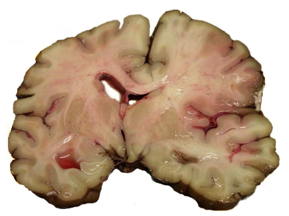
Image of MCA Stroke Brain Human, Courtesy of Marvin 101, CC BY-SA 3.0 <http://creativecommons.org/licenses/by-sa/3.0/>, via Wikimedia Commons
References
- GBD 2019 Stroke Collaborators. Global, regional, and national burden of stroke and its risk factors, 1990-2019: a systematic analysis for the Global Burden of Disease Study 2019. Lancet Neurol. 2021;20(10):795-820.
- Hacke W, Kaste M, Bluhmki E, et al.; ECASS Investigators. Thrombolysis with alteplase 3 to 4.5 hours after acute ischemic stroke. N Engl J Med. 2008;359(13):1317-1329.
- Parsons M, Spratt N, Bivard A, et al. A randomized trial of tenecteplase versus alteplase for acute ischemic stroke. N Engl J Med. 2012;366(12):1099-1107.
- Emberson J, Lees KR, Lyden P, et al. Effect of treatment delay, age, and stroke severity on the effects of intravenous thrombolysis with alteplase for acute ischaemic stroke: a meta-analysis of individual patient data from randomised trials. Lancet. 2014;384(9958):1929-1935.
- Berge E, Whiteley W, Audebert H, et al. European Stroke Organisation (ESO) guidelines on intravenous thrombolysis for acute ischaemic stroke. Eur Stroke J. 2021;6(1):I – LXII.
- Tong D, Reeves MJ, Hernandez AF, et al. Times from symptom onset to hospital arrival in the Get with the Guidelines–Stroke Program 2002 to 2009: temporal trends and implications: Temporal trends and implications. Stroke. 2012;43(7):1912-1917.
- Demeestere J, Wouters A, Christensen S, Lemmens R, Lansberg MG. Review of perfusion imaging in acute ischemic stroke: From time to tissue: From time to tissue. Stroke. 2020;51(3):1017-1024.
- Günkan A, Ferreira MY, Vilardo M, et al. Thrombolysis for Ischemic Stroke Beyond the 4.5-Hour Window: A Meta-Analysis of Randomized Clinical Trials. Stroke. Published online March 2025. doi:10.1161/STROKEAHA.124.048536
- Xiong Y, Campbell BCV, Schwamm LH, et al. Tenecteplase for ischemic stroke at 4.5 to 24 hours without thrombectomy. N Engl J Med. 2024;391(3):203-212.
- Zhou Y, He Y, Campbell BCV, et al. Alteplase for Acute Ischemic Stroke at 4.5 to 24 Hours: The HOPE Randomized Clinical Trial. JAMA. 2025;334(9). doi:10.1001/jama.2025.12063
- Jagolino?Cole AL, Dongarwar D, Aroor S, et al. Patients Residing in Rural Areas Transferred for Mechanical Thrombectomy Undergo Decreased Catheter?Based Treatment. Stroke: Vasc Interv Neurol. Published online March 2025. doi:10.1161/SVIN.124.001564
- Joubert J, Prentice LF, Moulin T, et al. Stroke in rural areas and small communities. Stroke. 2008;39(6). doi:10.1161/STROKEAHA.107.501643
- Mehta RH, Cox M, Smith EE, et al. Race/Ethnic differences in the risk of hemorrhagic complications among patients with ischemic stroke receiving thrombolytic therapy. Stroke. 2014;45(8):2263-2269.
- Rezai AR, D’Haese PF, Finomore V, et al. Ultrasound Blood–Brain Barrier Opening and Aducanumab in Alzheimer’s Disease. New Engl J Med. Published online January 4, 2024. doi:10.1056/NEJMoa2308719
- Tian W, Chu X, Tanzhu G, Zhou R. Optimal timing and sequence of combining stereotactic radiosurgery with immune checkpoint inhibitors in treating brain metastases: clinical evidence and mechanistic basis. J Transl Med. 2023;21(1):1-17.
- Fu AY, Bernstein K, Zhang J, et al. Outcomes of concurrent versus non-concurrent immune checkpoint inhibition with stereotactic radiosurgery for melanoma brain metastases. J Neurooncol. 2025;173(3):619-625.